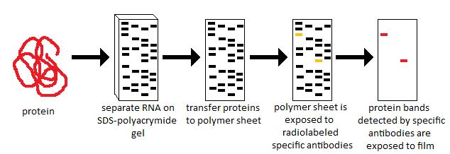
Western blotting is a widely used technique in molecular biology for detecting specific proteins in complex samples. Beyond simply visualizing proteins, researchers often need to quantify protein expression levels to compare samples, assess experimental effects, or validate findings. Accurate quantification requires careful selection of methods and proper normalization to ensure reproducible and meaningful results.
1. Densitometry-Based Quantification
Densitometry is the most common method for quantifying Western blot results. This technique involves measuring the intensity of protein bands from a digital image of the blot.
Workflow:
-
Capture a high-quality image of the blot using a digital imaging system or scanner.
-
Use software (e.g., ImageJ, Bio-Rad Image Lab) to define the area of each protein band.
-
Measure the integrated density or pixel intensity for each band.
-
Subtract background intensity to correct for non-specific signal.
-
Normalize the target protein signal to a loading control (such as β-actin or GAPDH).
Advantages: Simple, accessible, and compatible with chemiluminescent and fluorescent detection.
Limitations: Overexposed bands may exceed the linear detection range, leading to inaccurate quantification.
2. Fluorescent Western Blot Quantification
Fluorescent detection uses secondary antibodies conjugated to fluorescent dyes instead of enzymes. The signal is captured using a fluorescence scanner.
Workflow:
-
Incubate the blot with fluorescently labeled secondary antibodies.
-
Scan the blot with a fluorescence imaging system.
-
Measure band intensity for each protein using software.
-
Normalize signals to an internal loading control or total protein.
Advantages: High sensitivity, wide dynamic range, and allows simultaneous detection of multiple proteins on the same blot (multiplexing).
Limitations: Requires specialized imaging equipment and more expensive reagents than traditional chemiluminescence.
3. Chemiluminescent Quantification
Chemiluminescence involves enzyme-conjugated secondary antibodies that catalyze a light-emitting reaction. The emitted light is captured on X-ray film or a digital imaging system.
Workflow:
-
Apply chemiluminescent substrate to the blot.
-
Capture emitted light using film or a CCD camera.
-
Analyze band intensity using densitometry software.
-
Normalize against housekeeping how to perform a western blot or total protein levels.
Advantages: High sensitivity and widely available in most laboratories.
Limitations: Limited linear range if bands are overexposed; film-based detection is less quantitative than digital systems.
4. Normalization Techniques
Normalization is critical for accurate Western blot quantification:
-
Housekeeping Proteins: Target protein signals are divided by signals from stable proteins such as β-actin, GAPDH, or tubulin.
-
Total Protein Staining: Stains such as Ponceau S or Coomassie Blue measure total protein per lane, reducing variability caused by uneven loading.
-
Ratio Analysis: Express the amount of target protein relative to control samples to compare protein expression levels between conditions.
Proper normalization ensures that differences in band intensity reflect actual changes in protein levels rather than loading inconsistencies.
5. Advanced Quantification Approaches
Some researchers employ advanced methods to improve accuracy:
-
Non-linear regression analysis: Corrects for signal saturation in chemiluminescent blots.
-
Background subtraction algorithms: Improve accuracy in noisy images.
-
Multiplex fluorescence: Simultaneously quantifies multiple proteins and internal controls.
These methods enhance sensitivity and precision, especially in complex or quantitative studies.
6. Tips for Reliable Quantification
-
Ensure sample loading is within the linear detection range.
-
Avoid overexposure or underexposure when capturing images.
-
Always include replicates to account for biological and technical variability.
-
Validate that loading controls remain stable across experimental conditions.
-
Use consistent imaging and analysis parameters for reproducibility.
Conclusion
Western blot quantification methods provide researchers with the ability to measure protein levels accurately and reliably. Whether using densitometry, chemiluminescence, or fluorescence, the key to meaningful results lies in proper normalization, careful imaging, and consistent analysis. By combining careful experimental design with robust quantification strategies, Western blotting remains a powerful tool for studying protein expression, signaling pathways, and post-translational modifications in research and clinical applications.